ORIENTATION
http://www.springerlink.com/content/v8434087565413kk/fulltext.pdf?page=1
"Wellnhofer [4, 5] and Padian [6, 7], following von Zittel [8],described a system of fine structural fibers investing the [pterosaur] wing membrane, in a pattern similar to the orientation of the feather shafts of birds and the wing fingers of bats, both principal structural elements supporting the patagium and responsible for the transmission of aerodynamic force."
http://www.jstor.org/pss/2400656
The wing membrane was supported and controlled through a system of stiffened, intercalated fibers, which were oriented like the main structural elements in the wings of birds and bats.

MEMBRANE INERNAL STRUCTURE
http://www.ncbi.nlm.nih.gov/pmc/articles/PMC2842671/
"There are presently four competing models for the internal structure of the pterosaur wing membrane.Wellnhofer (1987) suggested that the actinofibrils were structural fibres embedded in the wing membrane.Pennycuick (1988) disagreed with this interpretation and regarded the ‘fibres’ as wrinkles caused by the inner elastic fibres contracting after the animal's death. In a review of pterosaur wing membrane, Padian & Rayner (1993) argued for the presence of structural fibres on the surface of the ventral part of the wing membrane, closely associated with the epidermis. Lastly, Tischlinger & Frey (2002) and Frey et al. (2003), based on a Rhamphorhynchus specimen from Solnhofen and an exceptionally well-preserved material from the Romualdo Formation (DGM 1475-R) of Brazil (Martill & Unwin 1989; Kellner 1996), interpreted the pterosaur plagiopatagium as consisting of five layers from dorsal to ventral: a thin and ‘hairless’ epidermis, a spongy subdermis, a layer of actinofibrils, a layer of dermal muscles and a vascular layer. Regarding the last model, it should be noted that Kellner (1996) regarded the soft tissue present in the specimen of the Romualdo Formation as closely associated with the body."
MATERIAL
http://en.wikipedia.org/wiki/Pterosaur
"The actual function of the actinofibrils is unknown, as is the exact material from which they were made. Depending on their exact composition (keratin, muscle, elastic structures, etc.), they may have been stiffening or strengthening agents in the outer part of the wing.[6] The wing membranes also contained a thin layer of muscle, fibrous tissue, and a unique, complex circulatory system of looping blood vessels.[7]"
"Since they [actinofibrils] were external, they were probably epidermal structures composed of keratin as in scales and feathers."
LAYERS
http://en.wikipedia.org/wiki/Pterosaur
"research has since shown that the wing membranes of pterosaurs were actually highly complex and dynamic structures suited to an active style of flight. First, the outer wings (from the wing to the elbow) were strengthened by closely spaced fibers called actinofibrils.[5] The actinofibrils themselves consisted of three distinct layers in the wing, forming a crisscross pattern when superimposed on one another."
http://rspb.royalsocietypublishing.org/content/early/2009/07/31/rspb.2009.0846.full
"The plagiopatagium can be divided into the distal, comparatively more rigid actinopagatium and a proximal, more tensile tenopatagium. The actinopatagium extends from the wing finger to the articulation between the humerus and the forearm, and shows the presence of at least three layers containing actinofibrils. In each layer, the actinofibrils are parallel to subparallel, but this direction diverges from layer to layer."
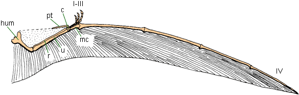
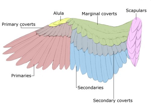
Modern bird:
UROPATAGIUM
http://rspb.royalsocietypublishing.org/content/early/2009/07/31/rspb.2009.0846.full
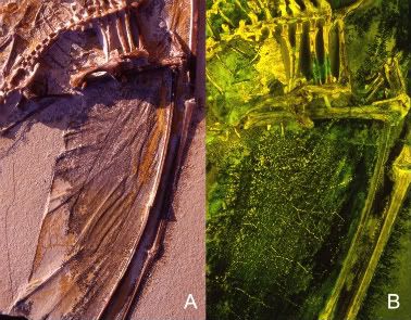
The uropatagium is not well preserved. Most of this membrane is encased in the bottom slab (plate I, fig. 2 in Wang et al. 2002), but traces can also be found in the top slab (figures 1 and 2). The shape of the uropatagium cannot be determined owing to the lack of a distinct posterior edge and the medially displaced feet. Two sets of fibres are observed: one running parallel to the longitudinal axis of the body and the second running perpendicular to the tibiae. This pattern is not as well developed as in the actinopatagium but, based on the thickness of the fibres, they are of the same nature as the actinofibrils. Integumental covering is also found.
No comments:
Post a Comment